Post-Concussion Dizziness: An Integrated Approach to Treatment and Recovery
Concussion injuries can trigger a multitude of symptoms, each unique and complex in its own right. The severity of these symptoms can vary considerably, ranging from barely noticeable to exceptionally severe.
Intriguingly, certain conditions—such as anxiety, ADHD, and a history of migraines or headaches—can heighten the likelihood of an individual experiencing more pronounced post-concussion symptoms.
While a vast spectrum of symptoms can manifest after a mild traumatic brain injury, certain symptoms tend to be more frequently reported among patients.
In this article, we’re going to concentrate on the specific symptom: “dizziness.” It’s a frequently encountered consequence of concussion injuries, and it’s imperative to understand its origins and implications.
We will discuss the primary causes, shed light on the processes involved in clinical decision-making, and explore the various available treatment options.
Clinical Decision Making:
PATIENT EXPERIENCING DIZZINESS
When a patient presents with a chief complaint of dizziness following a head injury, it’s crucial to properly triage and conduct a comprehensive clinical examination. Multiple factors can potentially contribute to the onset of dizziness, underscoring the importance of a detailed subjective history to inform the treatment pathway.
Whether in an outpatient clinic or hospital setting, proficiently interpreting and triaging the information provided to us as clinicians is paramount. It’s a skill that significantly influences our diagnostic precision and the subsequent care we render.
Symptoms, inherently subjective, can vary based on a patient’s unique experiences. Unlike tangible clinical signs we can directly observe or measure, symptoms hinge on patient self-reporting.
This characteristic can sometimes lead clinicians on a divergent path, perhaps resulting in a detour from optimal clinical decision-making. Such intricacies are particularly prominent when dealing with post-concussion dizziness.
Our primary goal when assessing a patient reporting dizziness is to identify the most likely root cause. The usual suspects in this investigation often include the vestibular system, the cervical spine, and the visual system.
In the ensuing discussion, we’ll discuss the sequential steps necessary for an effective evaluation and treatment of a patient experiencing post-concussion dizziness. Our aim is to ensure an encompassing understanding of this condition, leading to optimal patient care.
First Step:
RULE OUT RED FLAGS
The initial step in concussion care involves ruling out the necessity for additional imaging or the presence of a life-threatening injury. Notably, dizziness and vertigo may clinically signify severe injuries.
As outpatient clinicians, our primary role is to act as the first line of defense for our patients, escalating their care to advanced imaging if any component of our evaluation raises concern. By identifying and addressing these concerns, we can ensure patients receive the necessary care promptly.
Establishing a systematic protocol to rule out “red flags” or severe conditions is a vital first step. This system guarantees thoroughness in our approach and aids in delivering optimal care.
This protocol typically includes:
- Ruling out the need for CT scans or other advanced imaging
- Ruling out skull fractures
- Ruling out cervical spine fractures
- Ruling out cervical artery dissections
- Cranial Nerve Assessment
- Cerebellar Testing
- Upper and Lower limb Neurological examination
Awareness of the most common ‘red flags’ following a concussion is crucial. These include slurred speech, vomiting, unequal pupil sizes, prolonged loss of consciousness (greater than 30 minutes), escalating dizziness or loss of coordination, excessive drowsiness, difficulty waking the patient, and increased confusion or agitation.
Post-concussion dizziness is a commonly reported symptom, yet its presence necessitates a comprehensive and detailed evaluation due to the potential implication of a central lesion, particularly following an acute head injury. The pivotal question here is: does the patient require a computed tomography (CT) scan?
Well-established guidelines such as the New Orleans Criteria and the Canadian CT Head Rules serve as invaluable tools in this context. These criteria have a solid reputation for reliably determining patients who genuinely need further imaging, thereby minimizing the prevalence of unnecessary imaging procedures. This approach mitigates potential risks associated with CT imaging for those individuals where such measures are not medically indicated.
When dealing with pediatric patients, a slight deviation in the evaluation protocol is warranted. The Pediatric Emergency Care Applied Research Network (PECARN) rules are the gold standard for ascertaining the requirement for CT imaging in this demographic.
Another critical step involves excluding the possibility of skull and cervical spine fractures. The Canadian C-Spine Rules serve as a tool for identifying patients at a higher risk of fracture, thus indicating the necessity for further imaging studies.
When attempting to rule out the possibility of cervical artery dissections. The 2019 paper by Chaibi et al offers a detailed, systematic approach for mitigating this dilemma by evaluating the risk-benefit ratio. This invaluable resource includes a clinical decision tree, which can greatly enhance a clinician’s diagnostic accuracy.(1)
In every case of traumatic head injury, a comprehensive neurological examination should be performed. This examination should encompass a thorough assessment of cranial nerves, neurological evaluation of both upper and lower limbs, cerebellar testing, and the identification of any pathological reflexes.
When the patient presents with dizziness, this rigorous neurological evaluation serves to unveil additional insights into the patient’s condition, aiding in the exclusion of more severe underlying pathologies.
Vestibular Dysfunction
(BENIGN PAROXYSMAL POSITIONAL VERTIGO)
After ensuring the patient is not dealing with any serious pathologies, we can proceed to determine the cause of their reported symptom of dizziness.
Certain subjective descriptions such as “I experience dizziness when I change my head position” or “I have brief episodes of dizziness when I turn to lie on my side” could potentially indicate that the patient might be dealing with Benign Paroxysmal Positional Vertigo (BPPV).
Given the inherently subjective nature of dizziness, it is critical to differentiate whether the patient is actually experiencing vertigo or a different form of dizziness. While these terms are often used interchangeably, they possess distinct clinical meanings.
Vertigo is characterized by an illusion of self or environmental movement, typically described as “spinning” or “whirling.” Dizziness, on the other hand, refers to a sensation of unsteadiness or a feeling of motion within the head. Dizziness can be associated with fatigue, weakness, visual difficulties, or anxiety.
If the clinical assessment leans towards the presence of vertigo-like symptoms, BPPV can quickly be evaluated and potentially treated within the clinic setting. BPPV is a disorder of the inner ear, where the perception of our position in space is shaped through a sophisticated integration of various sensory inputs. One such mechanism is through otoliths, “the greek word for ear stones,” and semicircular canals.
Our inner ear houses these semicircular canals, filled with tiny crystals known as otoconia. As we change the position of our head, these crystals move accordingly, sending vital information to the brain about our spatial orientation. BPPV arises when this delicate system is disrupted.
The Dix-Hallpike test is the principal diagnostic tool for assessing the involvement of BPPV. Should the test yield positive results, the Epley’s maneuver is typically the first line of treatment. These methods predominantly target the posterior canals. If, however, the horizontal canals are implicated, the log roll maneuver can be performed.
Vestibular Ocular Motor Screening
(VOMS)
Once a patient has been definitively cleared of exhibiting vertigo-like symptoms attributed to BPPV, the next course of action typically involves a Vestibular Ocular Motor Screening (VOMS) assessment. This valuable tool aids the clinician in pinpointing various functional abnormalities that might have arisen following a concussion injury.
The VOMS assessment functions through a comparison of symptom scores recorded before and after the test. It is crafted in such a manner as to potentially provoke a patient’s symptoms, should there be significant involvement in the system being assessed. If a considerable increase in reported symptoms occurs after a particular test, it can provide valuable clues about the underlying causes of the post-concussion dizziness.
Composed of seven unique evaluations, the VOMS assessment meticulously probes specific functional deficits in the vestibular and visual systems. The individual tests within the VOMS assessment include:
- Smooth Pursuit
- Horizontal Saccades
- Vertical Saccades
- Convergence
- Horizontal Vestibulo-Ocular Reflex
- Vertical Vestibulo-Ocular Reflex
- Visual Motion Sensitivity Test
Each of these tests focuses on different aspects of the patient’s vestibular and visual performance, offering a comprehensive insight into their post-concussion health status.
Once the VOMS assessment is completed, the clinician can correlate its findings to the appropriate rehabilitation exercises. The primary goal of these exercises is to gradually expose the central nervous system (CNS) to varying levels of stimuli.
This methodology is rooted in the understanding that the perception and processing of afferent stimuli by the brain is a critical component in the dysfunction and subsequent treatment of visual and vestibular impairments.
The essence of these rehabilitative approaches lies in subjecting the CNS to controlled, graduated stimuli. To illustrate, if the patient demonstrates a dysfunction in smooth pursuits as per the VOMS assessment, their rehabilitation would be accordingly tailored. They would be given a series of exercises that incrementally raise the complexity of smooth pursuit tasks.
The bedrock of such rehabilitation lies in the principle of neuroplasticity, which implies that our brains are always adapting and evolving in response to the constant stream of stimuli. By providing the CNS with graded stimuli exposure, we can help it readjust to demanding real-life scenarios.
For more information on the VOMS, checkout our blog – The Vestibular Ocular Motor Screen: From concussion symptoms to concussion treatment- this test does it all
Will Provoking Symptoms Delay Recovery?
A significant aspect of the rehabilitation exercises derived from the VOMS assessment involves intentional slight provocation of the patient’s symptoms. If symptoms aren’t at least slightly provoked, then adequate recovery is unlikely to occur.
This concept is crucial to communicate to patients as it often raises apprehensions when there is a marked surge in symptoms. Patients should be advised to perform exercises to slight symptom provocation while staying below ‘significant symptom threshold’, which is defined as a 3 or more point increase on a 10 point scale from their starting point. Once this occurs, they should rest until their symptoms return to baseline, then repeat the exercise. As an example – if a patient has a resting overall symptom burden of 3 out of 10, then they should be aiming to raise their symptoms to a 5/10 and should stop and take a break at 6/10. .
The principles of neuroplasticity and adaptation work best with repetitive exercises, highlighting the importance of continual performance of these exercises throughout the day.
Symptoms are not indicative of brain health or recovery progress, and it’s crucial to push the perceived boundaries to stimulate the most change. This approach often yields the best results, leading to complete symptom relief.
Cervical Spine Involvement
DYSFUNCTIONS OF THE CERVICAL SPINE
The cervical spine plays a crucial role in contributing to symptoms of dizziness, given its high density of receptors located within the joints and musculature of the neck. These receptors provide essential proprioceptive feedback, informing the brain about our spatial positioning.
Dysfunctions in the cervical spine are known to manifest in the visual and vestibular systems. The mechanism involved is described as the cervico-ocular reflex and vestibular ocular reflex(2). This reflex is a normal pathway in our bodies that allow the cervical spine to relay proprioceptive information to the visual system.
Dysfunctions in the cervical spine such as tight musculature and joint restriction have been shown to cause visual and vestibular dysfunctions, tracking issues reflected in VOMS testing, and can lead to a range of symptoms including headaches, difficulty concentrating, memory issues, tinnitus, blurred vision and dizziness.(3)
The cervical spine, constantly absorbing information about our environment and conveying it to the central nervous system (CNS), can be disrupted following a head injury through two main mechanisms.
The first one is joint dysfunction. Each joint within our bodies, especially those within the cervical spine, contributes significantly to our sense of spatial orientation.
Following an injury, our bodies engage in protective or compensatory mechanisms that can lead to joint restrictions and muscle tightness. This has been observed to cause visual and vestibular disturbances in patients recovering from concussion injuries.
The visual and vestibular systems, along with the cervical spine, all relay information to the brain about our position in space. However, post-concussion, if the muscles in the cervical spine become tightened or shortened, they may begin to send inaccurate spatial information to the brain. This mismatch in information can lead to the patient’s perception of dizziness.
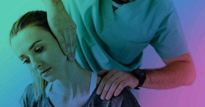
As clinicians, our responsibility lies in identifying and treating the dysfunction within the cervical spine. We do this through an array of manual therapy and rehabilitation techniques, aimed at rectifying the misinformation being conveyed to the brain and thus alleviating the perceived dizziness.
Determining The Involvement Of The Cervical Spine
FUNCTIONAL TESTING
Determining the involvement of the cervical spine in post-concussion dizziness can be achieved through various methods. An essential tool in this process is a clinical audit, which refers to a test or clinical finding, such as joint palpation, soft tissue palpation, or analysis of patient’s symptom severity. Having a reliable audit provides a benchmark to test against, leading to more effective and enduring treatment results.
The audits mentioned above—joint palpation and soft tissue palpation—are instrumental in assessing the cervical spine’s involvement. However, functional tests can offer additional insight and help refine the treatment methods employed by the clinician.
Several functional tests for cervicogenic dizziness include:
- Cervical Joint Position Error Test
- Bess-H Test
- Walking with Head Turns Test
- Smooth Pursuit Neck Torsion Test
Each of these tests can guide the clinician toward understanding the extent to which the cervical spine contributes to dizziness. The truly empowering aspect of these tests is their dual functionality: not only do they serve as diagnostic tools, they can also be used as part of the rehabilitation regimen. Paired with manual therapy techniques, these exercises can help facilitate the recovery process, offering an integrated approach to treatment and symptom relief.
Manual Therapy Of The Cervical Spine
JOINT AND SOFT TISSUE DYSFUNCTION
A particularly effective treatment for swiftly restoring proper joint function is the High-Velocity, Low-Amplitude (HVLA) joint manipulation. Often misunderstood, this technique doesn’t involve “realigning” bones in the traditional sense; instead, it targets restoring fluid movement and proper biomechanics to joints that have become restrained.
Skilled clinicians use palpation and observation to pinpoint these problematic joints, and through safe manipulation, they can restore their normal function. This process can significantly mitigate symptoms like dizziness.
Following a successful cervical manipulation, a window of improved joint function opens, paving the way for further manual therapy and rehabilitation techniques. Another critical element contributing to post-concussion dizziness is the presence of trigger points within constricted muscles. These trigger points are “hyperirritable spots” nestled within a taut band of skeletal muscle, which can arise due to various reasons such as improper joint mechanics, injury, inhibited stabilizing muscles, or flawed movement patterns — all of which are common post-concussion phenomena.
To address these trigger points, treatment may involve soft tissue therapy or dry needling. Aydin et al in 2018 performed a study on 55 women with self reported dizziness. The study found that the use of dry needling was effective at treating dizziness caused by cervical myofascial pain syndrome. The study also highlights the importance of an integrated model of care showing the combination of dry needling and therapeutic exercise as more effective than dry needling or therapeutic exercise alone (4).
Importance of an integrated technique
THE CERVICAL SPINE, VISUAL SYSTEM, AND VESTIBULAR SYSTEM
The cervical spine, visual system, and vestibular system all intricately interact with one another, reflecting a complex interplay of systems.
For effective treatment outcomes, it’s crucial to deploy a combination of techniques that address these connections. Treating one system in isolation can lead to limited improvement, as these systems collectively influence how you perceive your environment and reported symptoms. Studies have shown that focusing exclusively on vestibular training yields success in just 25% of patients(5). While effective for some, this relatively low success rate highlights the importance of adopting an integrated care approach to address all related systems for optimal results.
In Summary.
Addressing mild traumatic brain injuries requires swift medical attention at the initial stage of care. This immediate action is a cornerstone in the care of concussion injuries, playing a pivotal role in dramatically reducing the recovery period. While this discussion doesn’t cover all specialized considerations, it does underline the importance of paying special attention to symptoms like repeated vomiting, memory loss, skull fracture, impaired mental function, neurological problems, and brain swelling—all potential repercussions of head injuries.
Following incidents like a car accident or a head trauma sustained during contact sports, careful evaluation for signs of a skull fracture is necessary. Indicators such as “raccoon eyes” or a “Halo sign,” hinting at cerebrospinal fluid presence, can help guide the direction of treatment.
Identifying the cervical spine’s involvement is crucial and can aid the clinician in narrowing down the appropriate treatment methods. Vital clues in this context include reported motor vehicle accidents, post-traumatic headaches, repeated concussions, and balance problems.
The treatment of dizziness symptoms such as balance issues, difficulty paying attention, and double vision in patients with post-concussion syndrome requires a multi-modal approach. It’s worth noting that psychological factors can also contribute to dizziness. Common concerns often emerge, leading to questions like, “Should I worry about fatal brain swelling?”, “Does having had multiple concussions matter?”, or “Is there permanent damage to my brain cells?” Effective combat of such psychological concerns is best achieved through thorough education and reassurance.
Older adults, in particular, face an increased risk of a slow recovery following a serious injury. This emphasizes the importance of seeking medical assistance immediately after any injury occurs. All symptoms should be treated seriously, even if they appear mild at first or if the person initially seems symptom-free post-injury.
Patients with post-concussion syndrome can feel as if they don’t have normal brain function, but this is not the case. Patients perceive this notion because of chronic symptoms as a result of functional dysfunctions. Following the initial injury it’s crucial to recognize the signs and symptoms. If a second concussion occurs before the symptoms of a previous concussion have resolved, the risk of fatal brain swelling and permanent brain damage increases, as does the severity of symptoms. This highlights the need for thorough physical exams and meticulous medical treatment following a concussion injury, regardless of whether the cause was physical abuse, a blow to the head, or a blast injury from sports. It’s important to keep in mind that normal activities can exacerbate symptoms, and it’s advised to push through some discomfort and symptom exacerbation during the recovery period.
Ultimately, when treating post-concussion dizziness, it is crucial to adopt a systematic approach. The majority of patients presenting with post-concussion dizziness typically experience a combination of contributing factors. As healthcare providers, our primary responsibility during the initial visit is to rule out any serious underlying conditions, then to systematically uncover the underlying functional disturbances that may be causing the patient to experience dizziness. Clinical audits can prove extremely valuable in guiding the appropriate course of treatment.
Effectively managing mild traumatic brain injuries requires a comprehensive approach that addresses the involvement of all possible functional dysfunctions. The most common functional disturbances involve the cervical spine, visual and vestibular systems. The use of the VOMS assessment can be a great starting point for clinicians looking to determine the involvement of the visual and vestibular systems. Further evaluation and functional tests such as joint palpation, cervical joint position error test, and walking with head turn test can assist clinicians when determining the involvement of the cervical spine. To achieve optimal outcomes, a multimodal approach with the use of rehabilitation, manual therapy and lifestyle interventions should be used over isolated specific treatment interventions.
Denner Chiropractic & Performance | Charlotte, North Carolina | Concussion Specialist
At Denner Chiropractic & Performance, located in Charlotte North Carolina our concussion doctors specialize in the acute diagnosis, treatment, and rehabilitation of concussion injuries. Our rehab chiropractic care incorporates rehabilitation, joint manipulation, soft tissue, and dry needling to help you achieve pain-free movement in life and sports. Dr. Denner is certified through Complete Concussion Management (CCMI). CCMI is the world leader in evidence-based concussion treatment and rehabilitation. Denner Chiropractic & Performance has gone through the rigorous process of being a certified concussion clinic through CCMI. We are more than happy to discuss any concerns or questions you have about your condition or how we can help. Located on our main page or in our resource library tab is a sign-up for a free Discovery Call. During this time we will get to know you and your pain points. Let’s see if we are the right provider for you, schedule your Discovery Call today!
REFERENCES
- Chaibi A, Russell MB. A risk-benefit assessment strategy to exclude cervical artery dissection in spinal manual-therapy: a comprehensive review. Ann Med. 2019;51(2):118-127.
- Cheever K, Kawata K, Tierney R, Galgon A. Cervical Injury Assessments for Concussion Evaluation: A Review. J Athl Train. 2016;51(12):1037-1044.
- Schneider KJ, Meeuwisse WH, Nettel-Aguirre A, et al. Cervicovestibular rehabilitation in sport-related concussion: a randomized controlled trial. Br J Sports Med. 2014;48(17):1294-1298.
- Aydin T, Dernek B, Sentürk Ege T, Karan A, Aksoy C. The Effectiveness of Dry Needling and Exercise Therapy in Patients with Dizziness Caused By Cervical Myofascial Pain Syndrome; Prospective Randomized Clinical Study. Pain Med. 2019;20(1):153-160.
- Booth M, Powell J, McKeon P, Jennifer M, McKeon M. Vestibular Rehabilitation Therapy for Management of Concussion: A Critically Appraised Topic. International Journal of Athletic Therapy and Training. 2019;24(1):100-107

Denner Chiro Performance
Contact Me


